[ad_1]
In a current research printed within the journal Science Immunology, researchers profile the origins and subsequent differentiation of embryonic and fetal immune cells throughout human lung growth.
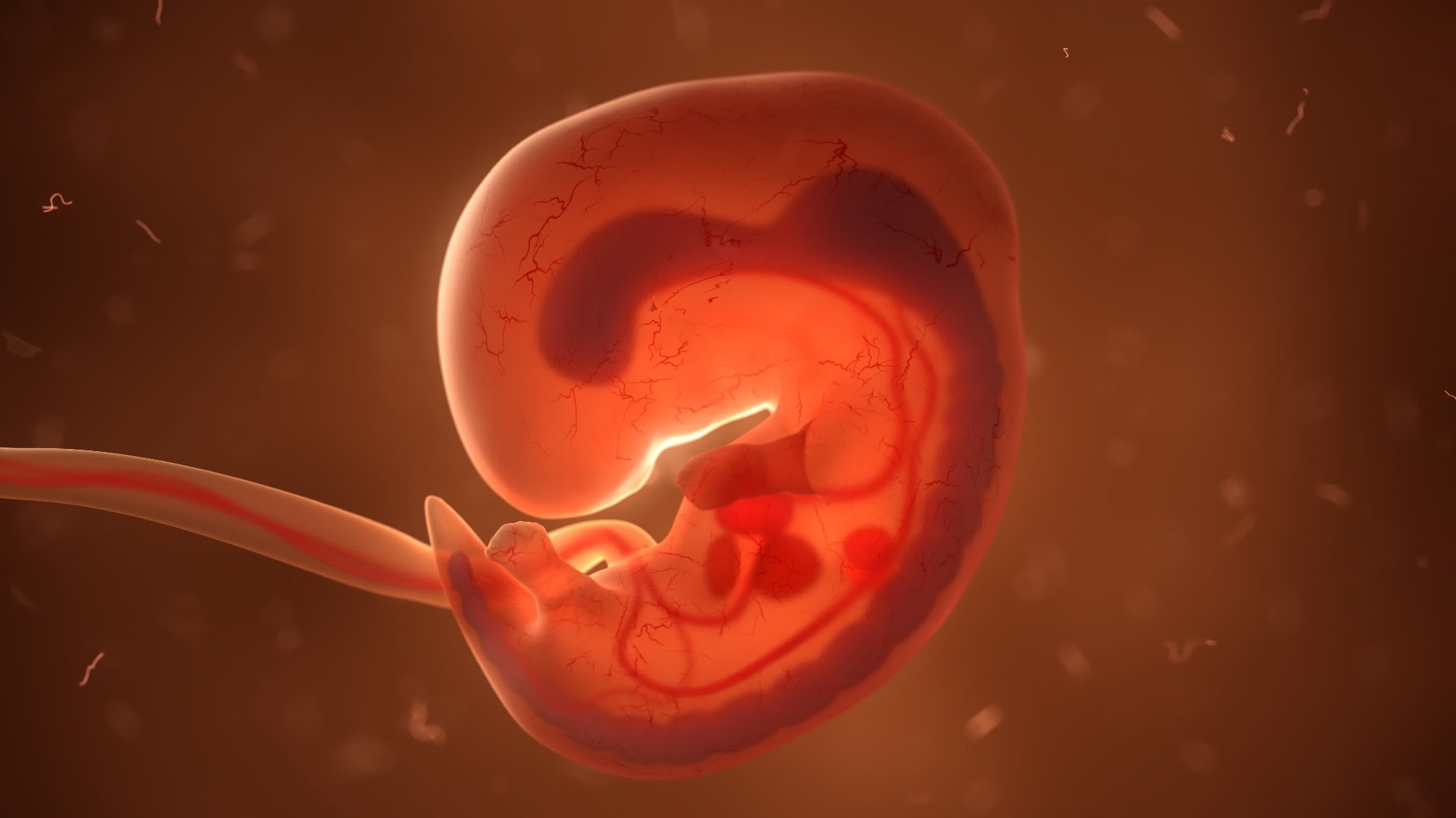
 Examine: Early human lung immune cell growth and its function in epithelial cell destiny. Picture Credit score: u3d / Shutterstock.com
What will we at the moment learn about fetal immune growth, and why is not that sufficient?
Earlier analysis has extensively documented the features of immune cells in regeneration, sustaining homeostasis, significantly within the gut and testis, and somatic tissue growth. Research have aimed to elucidate the construction, subtypes, and performance of epithelial and mesenchymal cells; nonetheless, a niche exists in researchers’ understanding of the processes and purposeful roles of lung immune cells.
Immune cells are among the most important cells liable for an toddler’s survival from beginning onwards. Given the defenses supplied by lung-associated mucosal immune cells in opposition to airborne pathogens and inhaled toxins, the present dearth of literature on the topic is shocking. A doable rationalization for this hole within the literature could also be as a result of advanced nature of cell differentiation throughout embryonic growth and the historic lack of strategies able to safely tracing these differentiations all through being pregnant.
A crucial query that continues to be unanswered is whether or not immune cells might need features over and above protection – may they modulate or in any other case affect the event of the tissues whereby they reside? Answering this and related questions pertaining to human lung growth at each mobile and molecular ranges could consequence within the genesis of novel medical interventions designed to restore and regenerate lungs, thereby affording hundreds of thousands and even a whole bunch of hundreds of thousands of sufferers an alternative choice to lung transplantation.
Earlier analysis has characterised human lung developmental morphology and categorised the method into 5 temporally overlapping phases. These encompass the embryonic stage between 4 and 7 weeks submit conception (pcw), the pseudoglandular stage between 5 and 17 pcw, the canalicular stage between 16 and 26 pcw, the saccular stage between 24 and 38 pcw, and the alveolar stage from 36 pcw to 21 years of age.
The primary three phases, particularly between 5 and 22 pcw, signify the least understood interval of lung growth regardless of their collectively protecting your complete evolution of epithelial stem cells into virtually purposeful lungs.
Concerning the research
The current research aimed to judge the temporal development of the fetal immune system and elucidate its potential function in modulating embryonic lung growth. Human fetal and embryonic samples have been acquired from the Human Developmental Biology Useful resource (HDBR) Joint MRC/Wellcome Belief grant.
Pregnancies terminated between 5 and 22 pcw have been used to acquire contemporary lung tissue with written consent from donors. Karyotypic evaluation was carried out to make sure that included samples have been free from genetic abnormalities and represented ‘typical’ human embryonic progress.
Immunohistochemistry (IHC) of lung tissues was used to validate immune cell varieties and amount throughout the primary three phases of embryonic lung growth. IHC evaluation additional contributed to evaluating the areas of noticed immune cells and variations therein from 5 to 22 pcw.
To enhance the accuracy and reliability of immune cell quantification, three-dimensional (3D) quantification utilizing confocal microscopy adopted by Imaris software program analyses was carried out. Computed 3D photos have been in comparison with 2D photos at each time level throughout the research length.
Lung tissue digestion adopted by movement cytometry and fluorescence-activated cell sorting (FACS) have been used to validate IHC quantification estimates, elucidate relative proportions of CD3+, CD4+, CD8+, and regulatory T cells (Tregs) as a proportion of CD45+ populations for a similar developmental stage, and kind CD45+ cells as a precursor to single-cell ribonucleic acid (RNA) sequencing (scRNA-sq).
Furthermore, scRNA-sq served the twin goal of validating movement cytometry outcomes and the molecular characterization of immune cells throughout samples. Mobile indexing of transcriptomes and epitopes sequencing (CITE-seq) was moreover employed to enhance the decision of the outcomes.
Human embryonic lung organoids have been additionally used for purposeful characterization experiments, together with cytokine therapies, twin Suppressor of Moms In opposition to Decapentaplegic (SMAD) transcription assays, and macrophage, dendritic cell (DC) tradition, and cytokine arrays.
All obtained information have been topic to statistical analyses consisting of one-way Evaluation of Variance (ANOVA), unpaired two-tailed t-tests, residual most probability evaluation (REML), and Tukey’s submit hoc multiple-comparison take a look at.
Examine findings
The scRNA-seq, IHC, and purposeful organoid assays revealed that immune cell populations various considerably throughout fetal developmental phases. Progenitor and innate immune cells, together with myeloid, innate lymphoid (ILC), and pure killer (NK) cells, predominated in early developmental phases however have been step by step changed by T- and B-lymphocytes. CD45+ cells have been almost ubiquitous throughout developmental phases and in all lung-associated tissue areas; nonetheless, their relative amount various throughout time and site.
Molecular characterization of immune cells revealed 77,559 transcriptomic profiles, 61,757 of that are novel to science. Annotation of those profiles adopted by clustering analyses revealed 59 clusters consultant of all identified immune cell classes. Analyses of the progenitors of those classes resulted within the discovery of unexpectedly excessive ILC- and early lymphoid progenitor (ELPs) densities.
Taken collectively, these outcomes counsel that immune cells comply with a biphasic sample throughout fetal growth and a spike in abundance throughout eight and 20 pcw. Quantitivate polymerase chain response (qPCR) evaluations counsel that the 20 pcw peak could also be partially on account of vascular maturation.
B-cell maturation in lungs was revealed for the primary time utilizing IHC and single-molecule fluorescence in situ hybridization (smFISH) assays. This added to a rising physique of proof that the bone marrow is just not the only supply of mature B-cells, which is opposite to earlier scientific beliefs.
Immunoglobulin (Ig) isotype expression in tandem with clonal enlargement assays revealed that, throughout growth, lung mesenchyme and epithelium help B-cell homeostasis via the secretion of modulatory chemokines, together with CCL28.
The collective output of transcriptomic and cytokine assays revealed the advanced interactions of a number of immune cell-secreted cytokines, which, in flip, have been functionally validated to have an effect on epithelial cell differentiation. These outcomes counsel that immune cells serve a twin goal of protection and lung growth throughout fetal growth, thereby confirming earlier hypotheses.
Conclusions
Within the current research, researchers mixed cutting-edge transcriptomic analyses with IHC to elucidate immune cells’ structural and purposeful roles throughout fetal and embryonic growth. Evaluations of fetal immune cells through the 5 to 33 pcw interval revealed that full B-cell maturation happens in embryonic lungs, which contests the prevalent perception that the bone marrow is the only supply of mature B-cell populations.
Interleukin-1 beta (IL-1β) was discovered to be extensively produced by broadly dispersed myeloid cells. IL-1β, in flip, was discovered to modulate and promote epithelial stem cell differentiation, highlighting the twin function of immune cells in each protection and lung epithelial growth.
Collectively, these findings present an immune atlas of growing human lungs and counsel a job for fetal immune cells in guiding growth of the lung epithelium.”
Journal reference:
- Barnes, J. L., Yoshida, M., He, P., et al. (2023). Early human lung immune cell growth and its function in epithelial cell destiny. Science Immunology. doi:10.1126/sciimmunol.adf9988
[ad_2]